
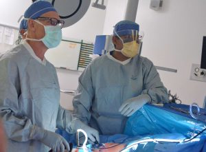
SHOULDER ARTHROSCOPY
Arthroscopy is a minimally invasive surgical and diagnostic procedure performed for joint problems. Shoulder arthroscopy (αρθροσκόπηση ώμου) is performed with a pencil-sized instrument called an arthroscope. The arthroscope consists of a light system and a camera to project images onto a computer screen. So your surgeon can see the surgical site. We use arthroscopy to treat diseases and injuries that affect the bones, cartilage and muscles of the shoulder joint.
Disease overview: The shoulder joint is like a ball-and-socket joint, where the head of the humerus (upper arm bone) articulates with the glenoid of the scapula (shoulder blade) called the glenoid. Cartilage covers the two articulating surfaces of the bones are. It prevents friction between the moving bones allowing smooth movement.
Ligaments and tendons around the bear joint give quality and soundness to the joint. Injuries and diseases to the bones or soft tissues of the shoulder joint can make it unstable. This leads to pain, swelling, and reduced mobility.
Indications
Shoulder arthroscopy demonstrates to treat the taking after bear conditions when preservationist treatment. Pharmaceutical and treatment, falls flat to soothe torment and inability:
– Rotator cuff tear.
– Stiff shoulder or frozen shoulder joint.
– Shoulder Instability. This occurs when the head of the upper arm bone slips out of the socket of the glenoid fossa of the shoulder blade either through injury or overuse.
– Biceps rupture occurs when tendons attach the biceps muscle to the shoulder or elbow tears.
– Damaged cartilage or ligaments.
– Bone spurs or bony projections.
– Arthritis of the clavicle.
Process
Your surgeon performs a shoulder arthroscopy under general or regional anesthesia. You may place yourself lying on your side with your arm to support or sit in a semi-recumbent position. Surgeon infuses sterile liquid is into the bear joint to extend the surgical region. This way your specialist contains a clear see of the harm and room to work.
Hence a button-sized gap goes within the bear and the arthroscope (αρθροσκόπηση ώμου) embedds. Your surgeon can see images seen at the camera of an arthroscope on a monitor. Surgical instruments go into the joint (αρθροσκοπηση γονατος) through separate small holes. At this time they remove and repair damage to the joint. After surgery, doctor expels the rebellious and closes the cuts with fastens or little sterile gauze strips.
Postoperative care: After surgery, small surgical wounds take a few days to heal and the surgical dressing is replaced with simple band-aids. Recuperation time depends on the sort and degree of the performance of the strategy. They prescribe you pain medications to keep you comfortable. You place the arm on the shoulder that had the problem in a sling for a short time. Afterwards they recommend you physical therapy to improve shoulder mobility and strength after surgery.
Advantage
The preferences of arthroscopy (Αρθροσκόπηση Ώμου) when you compare them to open surgery with a huge entry point include:
– Less pain.
– Fewer complications
– Shorter hospital stay.
– Shorter recovery.
Risks and complications: Complications of shoulder arthroscopy – αρθροσκόπηση ώμου include infection, bleeding, damage to nearby nerves or blood vessels, or delayed healing after surgery. In certain cases, stiffness of the shoulder joint can occur after surgery.
It is important to actively participate in your physical therapy to prevent this from happening.
What is a Sports Injury?
Within sports injuries we can find:
Acromioclavicular Dislocation: The history often points to direct trauma to the shoulder with the arm adducted or hanging along the body. Clinically, impotence is partial. There are four grades in the acromioclavicular joint injury, the same ones that we apply for the management of each case.
In grades 0 and 1, the treatment is functional, the arm keeps in a sling for a period of 2 to 3 weeks. Treatment in grade 3 is the most controversial between supporters of functional treatment and those of surgical treatment. Such as shoulder arthroscopy – αρθροσκόπηση ώμου.
Sternoclavicular dislocation
The displacement can be anterior or posterior, but the weakness of the anterior sternoclavicular ligament (ACL) with respect to the posterior sternoclavicular ligament (PSCL) explains why in most cases, the displacement is anterior.
Treatment for anterior sternoclavicular dislocations is functional, consisting of a short period of arm immobilization in a sling until pain subsides; the only sequel that can remain is aesthetic; or orthopedic. A support will be made by means of a bandage with the elbow close to the body for 6 weeks.
FROZEN SHOULDER
It consists of the loss of passive mobility of the shoulder, especially external rotation, which is accompanied by diffuse pain and functional limitation.Its course is deceptive, dynamic and, in most cases, self-limiting. It occurs more frequently between 40 and 70 years; It is more common in women and patients with thyroid disorders or diabetes.
The goal of treatment is to restore joint mobility and functionality, as well as improve pain through the use of anti-inflammatories, steroids, physical therapy, and sometimes closed manipulation under anesthesia or capsular release through arthroscopy. Shoulder mobilization exercises are indicated from the onset of capsulitis and in the immediate postoperative period. Mobilization under anesthesia and arthroscopic capsular release are considered safe and effective methods that allow patients to have better mobility and pain control.
Posterior Dislocation
It is not very common (1 to 4% of shoulder dislocations) and often goes unnoticed. Often the mechanism is indirect, by performing a forced internal rotation. It usually occurs in adults, especially men. The pathognomonic sign is the presence of irreducible internal rotation leading to the impossibility of external rotation (with the arm extended, the patient is unable to turn the palm of the hand upwards).
The treatment corresponds to performing an emergency reduction, normally with general anesthesia, by means of traction, adduction and external rotation maneuvers. Immobilization is performed with the elbow close to the body, in neutral rotation and for 3 weeks. In the case of a misdiagnosed old dislocation, it is not uncommon to resort to surgical reduction.
Posterior Dislocation
It is not very common (1 to 4% of shoulder dislocations) and often goes unnoticed. Often the mechanism is indirect, by performing a forced internal rotation. It usually occurs in adults, especially men. The pathognomonic sign is the presence of irreducible internal rotation leading to the impossibility of external rotation (with the arm extended, the patient is unable to turn the palm of the hand upwards). The treatment corresponds to performing an emergency reduction, normally with general anesthesia, by means of traction, adduction and external rotation maneuvers. Immobilization is performed with the elbow close to the body, in neutral rotation and for 3 weeks. In the case of a misdiagnosed old dislocation, it is not uncommon to resort to surgical reduction.
INSTABILITY
Bear precariousness happens when the head of the arm bone is constrained out of the bear attachment. This may happen as a result of an unanticipated harm or from overuse.
Shoulder dislocations can be partial, when the ball of the arm only partially protrudes from the socket. This is called a subluxation. A total disengagement implies that the spheroidal portion comes totally out of the cavity. Once the tendons, ligaments, and muscles around the bear ended up free or torn, separations can happen more than once. Recurrent dislocations, which can be partial or complete, cause pain and instability when you raise your arm or move it away from your body. Repeated episodes of subluxations or dislocations lead to an increased risk of developing osteoarthritis in the joint.


